By Nancy Frishkorn BA, CHCP (reposted from September 10, 2013)
If you own horses, chances are good that at some point either you or someone you know spent many hours tending to an abscess. An abscess is collection of pus in an area of the body (in this case the hoof capsule) that causes severe pain and swelling due to the body’s immune system’s attempt to fight off the infection. This pus is actually excess white blood cells and tissue (living and dead), fluid, bacteria and other foreign substances. The white cells are the body’s natural defense to infection that release destructive components after identifying and binding with bacteria. Their purpose is to “kill” the harmful bacteria, but in the process healthy tissues are also damaged. In the hoof, this damage most often occurs in the laminae and bony structure within; in other words, if not treated, the coffin bone itself begins to degenerate and weaken, causing small pieces to break away. As part of the inflammation response, more white cells are sent to the site to remove the damaged tissue (the clean-up crew) which actually creates even more inflammation and subsequently more pain. The pieces of broken and damaged tissue are not distinguished by the body and the natural immune system subsequently treats them as foreign objects; hence, the system treats the bone pieces as “foreign objects” – these are what are known as sequestrum.
This is the story of Colt, a beautiful gelding purchased by Carla (Pittsburgh Pet Connections CEO) who had poor hoof care before she found him. There are some individuals who believe the hooves can go months without trimming, and others who feel they can trim themselves despite the fact that they have had no training or poor training at best. Colt was one such victim of circumstance, and he came into Carla’s love and devotion in need of immediate attention. His hooves were long and imbalanced, and after two trims he was still experiencing intermittent lameness. Local vets were called and his abscess was opened, but they continued to fester despite many hours of soaking, draining and treatments with drawing salve. After seeing no improvement, it was decided he needed to seek clinical attention for a second opinion and x-rays.
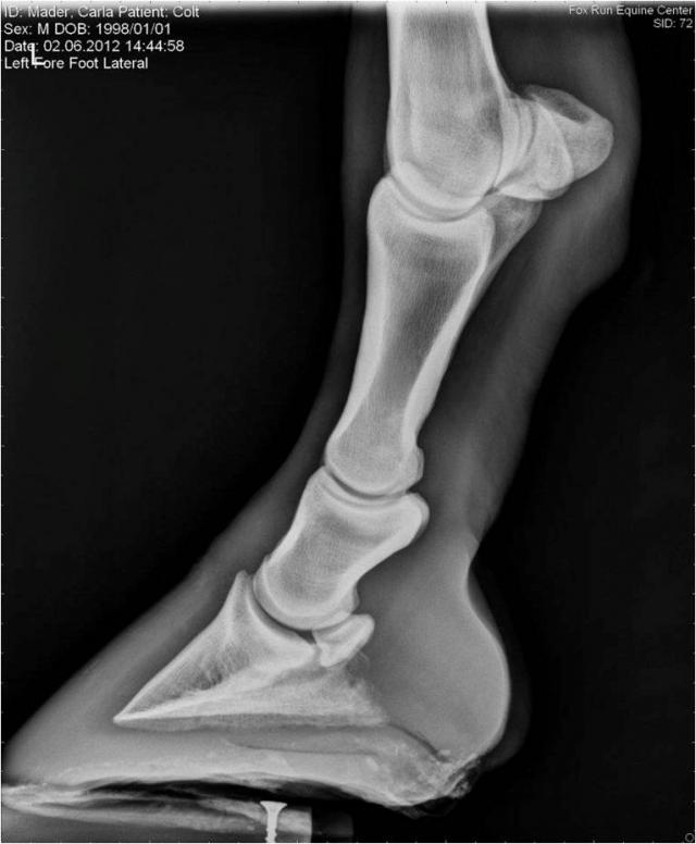
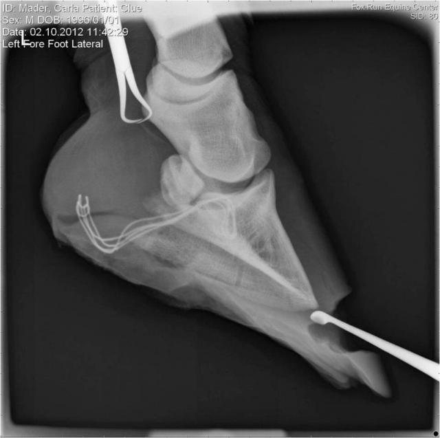
Colt was sent to Fox Run Equine Center where Dr. Brian Burks DVM diagnosed a lateral sequestrum on Colt’s left front hoof. This first picture shows Colt’s tract on film; you can see some lines coming from the side of the hoof draining down by the back of the heel.
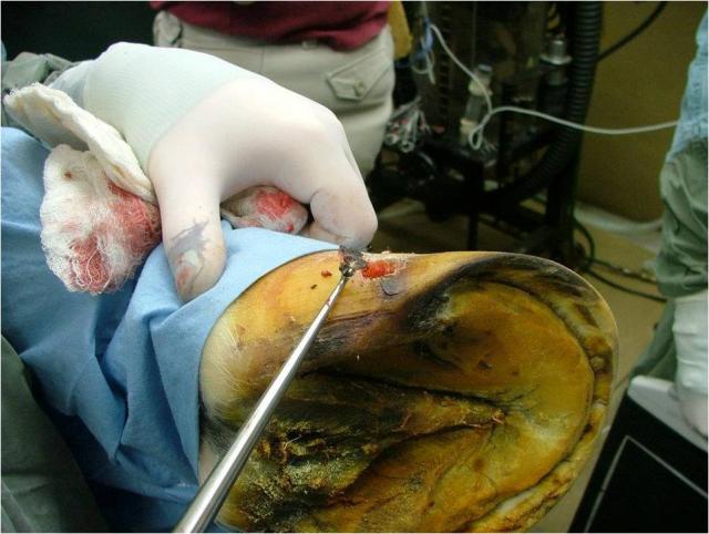
This is the site that had been opened from the outside bar (hoof wall beside the frog) but never drained out completely. Inside, there is a piece of broken bone that was damaged due the accumulation of pus for a long period of time. Dr. Burks used a dremel tool to drill a small hole into the quarter (side of the hoof wall) to remove this sequestrum. The second picture shows the piece of bone being removed and just how small the piece of bone was; its removal was imperative for Colt’s recovery.
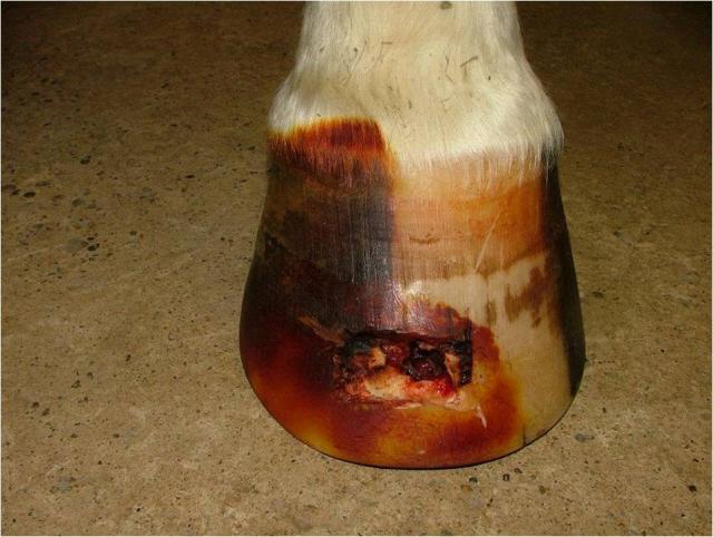
The third picture is a shot of this same area after surgery, the quarter area grew out within three months with daily packing with betadine and Sliver Sulfadiazine.
Before the surgery, Dr. Burks scraped out all the hard laminae from the bottom of the hoof to ensure there would be no residual bacteria’s invading the capsule that could potentially cause reinfection of the hoof. His intuitions served him well when it was discovered that the very tip of P3 (coffin bone) was extremely brittle. He concluded that this was damaged a long time ago from old abscessing that had caused this area to weaken and nearly break away. By making another “window” in the hoof wall, Dr. Burks was able to preserve most of the wall structure and remove this weakened area as well. He commented to me that the tip “fell away” when he merely touched it with his forceps, so it too was removed and needed packing until it grew out. This fourth picture shows the actual procedure during surgery when the forceps were inserted into the toe wall to remove the sequestrum.
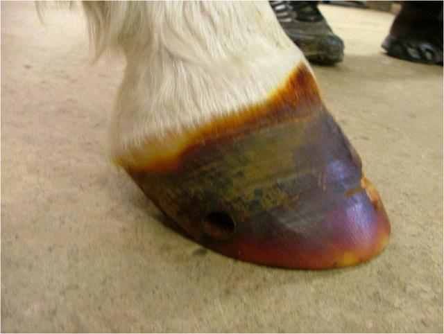
I’ve worked with many vets over the years, but I’ve never met one quite as thorough and open minded as Dr. Burks. The traditional protocol for any respective procedure is hospital plates (wide aluminum shoes) that stay on for many months to support the hoof during healing. Because Burks took such care to make minimally invasive openings for removal, Colt was left with adequate hoof wall for support. Carla was adamant in keeping Colt as natural as possible, meaning she wanted him to remain barefoot, and he respected her wishes. I was called to meet with Burks about follow up hoof care and we mutually agreed he could remain in a hoof boot that would not only support his hoof, but also provide better coverage for the opened areas that needed daily treatments. This last picture shows Colt’s open toe area five days after surgery when he was taken out of wraps and placed in a hoof boot.
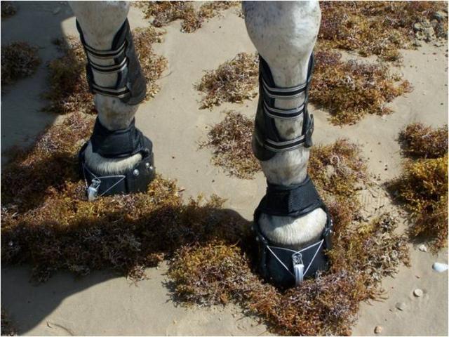
Treating a hoof injury is difficult on the owner as well as the horse. Carla was going to need a boot that would not only cover the entire hoof wall, but also one that could be easily removed and strong enough to withstand several months of continuous wear. Colt was rather stubborn about lifting the hoof for his daily treatment, so ease of application was an absolute necessity. I am familiar with several boots, but the best choice for this situation called for durability, full support and easy removal as well so that no further damage would occur. I could think of only one boot that would serve her purpose, and one that she would be able to keep for years to come in case she ever needed them again – the Easyboot Rx.
From March to mid-May Colt wore his boots day and night. He was sound at a walk almost immediately after the surgery and because he had a boot he was able to get turnout in the arena and a small paddock every day. We actually booted both front hooves to make sure he wasn’t off balance on the front and this kept him sound while simultaneously avoiding any shoulder pressure or further injury. Carla made sure that his hooves were kept as dry as possible to avoid any rubbing due to excess moisture or sweat by removing them daily for treatments and drying the back of the hoof before replacing it. This movement helped facilitate the healing process and by the end of May the entire wall had grown out completely with no further problems. Within a month Colt was even able to do short rides wearing hoof boots and today he is doing very well. He has not had an abscess in nearly a year and his soles are tough because he has relocated to a facility that enables full turnout and natural wear. Carla has since purchased a pair of Easyboot Trail boots for long rides, and we are grateful to not only EasyCare for their supreme products, but also to Dr. Burks for his open-minded approach to natural horse keeping. Thanks to Carla, Colt has a wonderful life and his hoof issues are no longer…he is happy, healthy, and sound.
– Nancy Frishkorn BA, CHCP


