


In previous blogs I’ve written about my basic hoof guidelines:
- 3-8 degree palmar P3 angle: the angle of the bottom of the coffin bone in relation to the ground.
- 50/50 base of support from toe to heel around the center of rotation of the hoof capsule.
- Capsular and phalangeal alignment, with a straight hoof-pastern axis.
- Minimizing flare and distortion in the hoof capsule.
And I’ve even touched upon some of the reasons why those guidelines have become central for me:
https://blog.easycareinc.com/blog/hoof-love-not-war/is-your-horse-really-a-goat
However, at the heart of those guidelines and the work I do with each horse’s foot, is the consideration of circulation. The circulation of the horse’s foot is critical to nourishing the tissues as well as being an integral part of the energy, shock and heat management system of the foot.
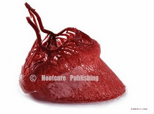
The circulation of the horse’s foot is just fascinating. This is a platinate corrosion cast of the blood vessels of the horse’s foot by Dr Christoph von Horst of HC Biovision. Note how intricate the vessels appear:
Dr. Bob Bowker is one of the first researchers I heard years ago discuss the importance of specific trimming ideas designed to support healthy circulation. He referenced a 3-5 degree Palmar P3 Angle would allow for appropriate load on the back of the foot, facilitating good circulation and the growth of healthy soft tissue.
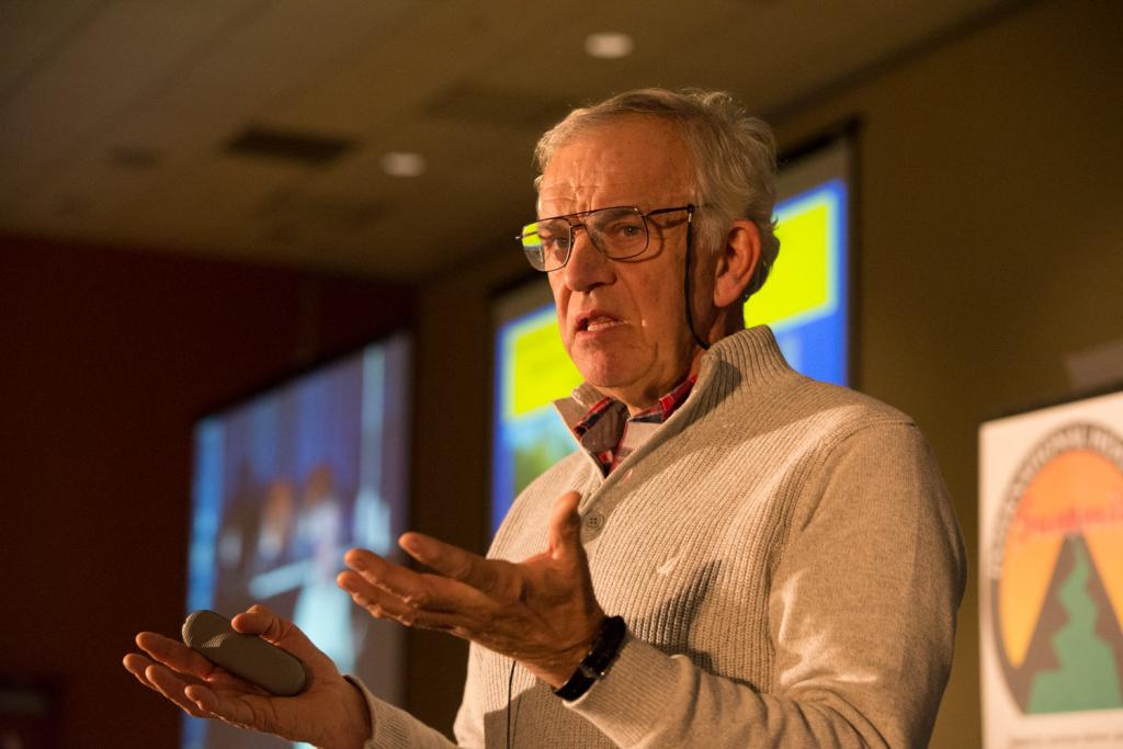
Before Dr Bowker’s latest research, recently presented at the 2015 International Hoof-Care Summit, the smallest vessels we could see were 500 microns. Now because of his work we can appreciate the blood vessels down to 1 micron in diameter.
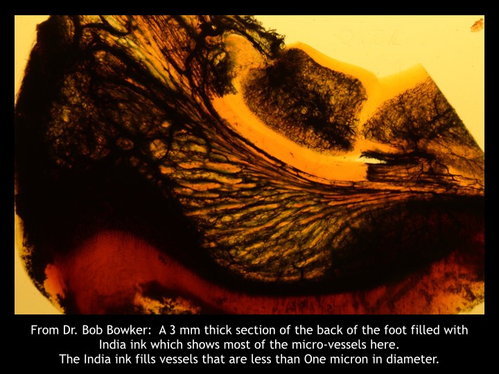
To give you an idea of how small a micron is: 1 micron = .00004 inches or .001mm. A human hair is 50-80 microns, and 10 microns is the smallest size visible to the human eye.
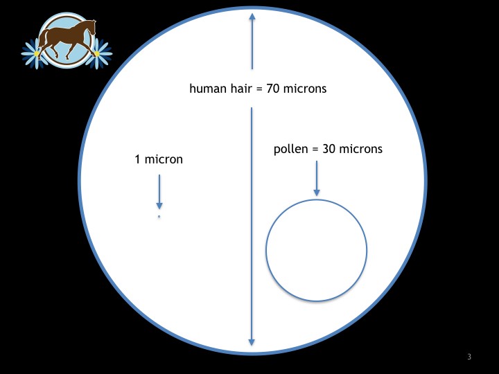
So these blood vessels are very small and highly influenced by load and pressure.

We can experience how these blood vessels can be influenced by pressure by pressing and removing our thumb on our forearm and watching the color (blood) come back to the area, similar to checking capillary refill on the gums of a horse. Therefore the arrangement of internal and external anatomy of the foot and how the horse loads it’s feet similarly influences the circulation of the foot. Through our hoof care work, we also greatly influence how the horse loads it’s foot.
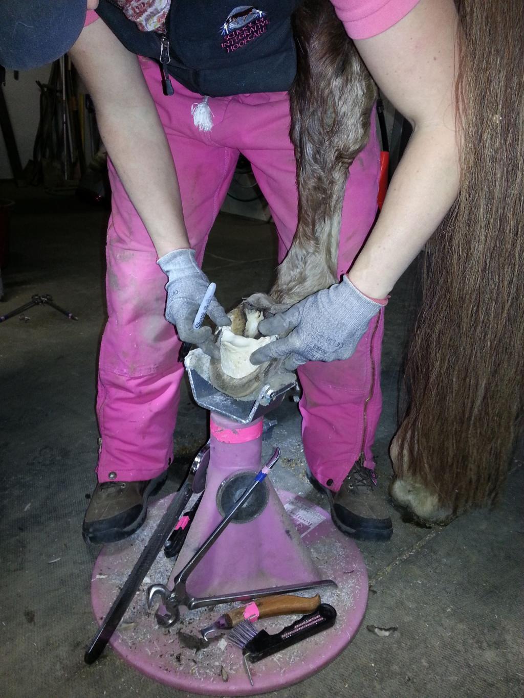
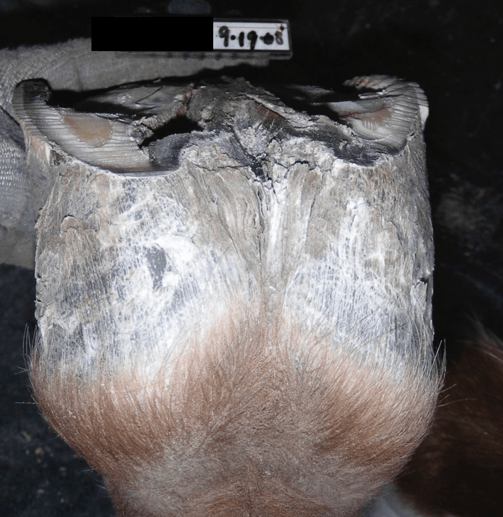
One of the most impactful ways I experience this dynamic was in 2008 when I was asked to help a horse who had been chronically foundered for quite a while. While he had been receiving excellent farrier care, he was quite painful:
His situation was complicated by lack of any growth at the dorsal wall despite a controlled metabolic situation with an excellent diet. With the previous farrier’s blessing I went to work on this horse and found that no matter how much I tried to realign the hoof capsule with the internal anatomy, I ran into the same struggles: lack of growth at the coronary band at the toe, and an inability to load his heel if I trimmed his heel down. In addition to those problems, he had a thin, convex sole.
Then I read an article in The Horse which summarized the work of Lorenzo D’Arpe, DVM, PhD regarding venograms and how different hoof angles applied to the same foot affect blood flow: http://www.thehorse.com/articles/18491/laminitis-coming-out-of-the-dark-bluegrass-laminitis-symposium
The part that really stood out to me was: “…changing the palmar angle modifies the valscularization (blood supply) of the foot.” I hypothesized that the reason this horse was stuck was in large part due to how little improvement could be made to his phalangeal and capsular alignment over time. For more information about these alignment problems, see: https://blog.easycareinc.com/blog/hoof-love-not-war/rehabilitation-of-the-insulin-resistant-foundered-horse-dhf-style
The veterinarian helping us with this horse, Dr. Mark Donaldson of Unionville Equine in Oxford, PA, performed a venogram so we could observe the circulation of his foot. Here is the venogram showing the compromised blood flow of this foot at that time.
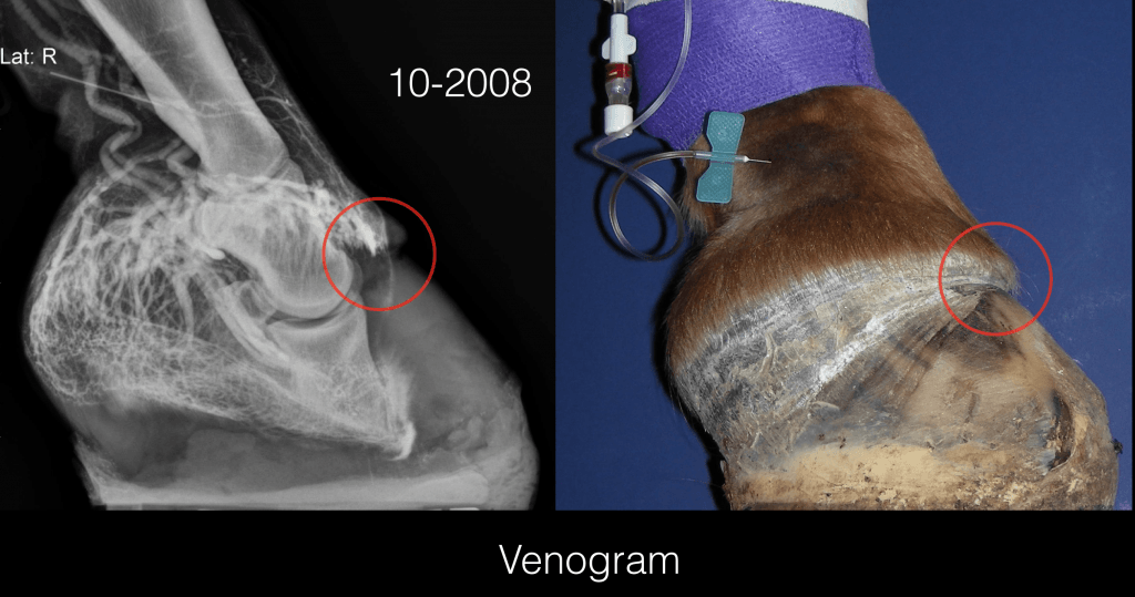
So far my hypothesis was correct. This venogram showed lack of perfusion of the blood supply past the coronary band and along the dorsal surface of P3. We decided more drastic intervention was necessary to try to make a positive change for this horse. Dr. Donaldson and horse owner elected to do a deep digital flexor tenotomy in an effort to allow me to realign the hoof capsule and internal structures. We weren’t sure how much of the circulation to the front of the foot could be re-established, however we believed was the best chance we had.
After the tenotomy we saw immediate improvement in the horse’s comfort, and finally some growth of wall at the coronary band at the toe. On the follow-up venogram we saw significant improvement in the circulation. Here is a venogram 10 weeks after the tenotomy:
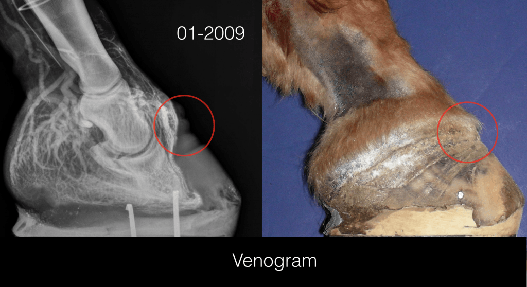
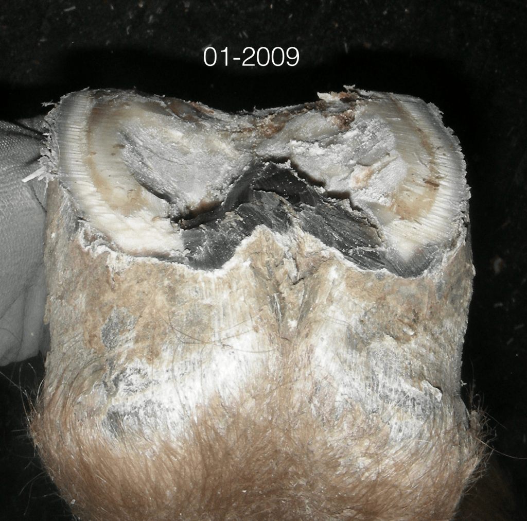
We had significantly better circulation. The new growth wasn’t pretty but at least we were getting something. And the best part was horse’s comfort level continued to improve. Here is his foot and the growth nine months post tenotomy:
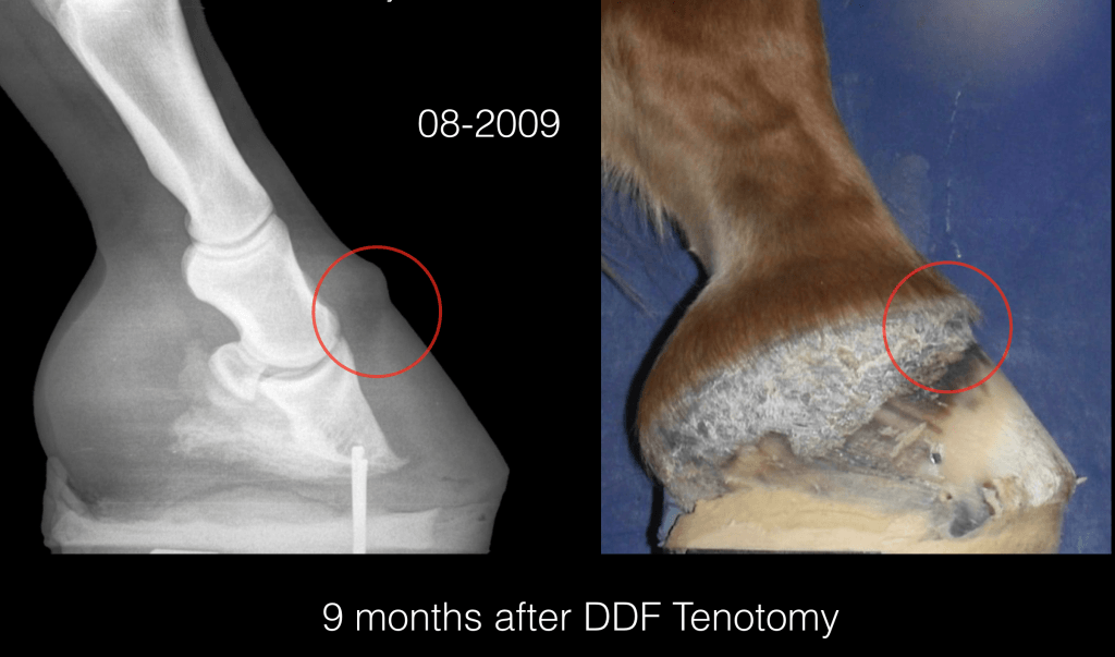
Even his sole was no longer convex:
Working on this horse and thinking about load and circulation pushed me to get creative and develop a strategy with the horse’s team that led to his soundness and a much healthier foot. When you think about the horse’s foot, I encourage you to consider circulation as well.


